
Sidoryak N.G., Putiy Y.V., Rozova E.V. INVESTIGATION OF STRUCTURAL-FUNCTIONAL CHANGES AND FEATURES OF GENES - CANDIDATE EXPRESSION IN TISSUES OF MEDULLA OBLONGATA AND STRIATUM UNDER EXPERIMENTAL PARKINSONISM
Участников: 3
Интернет-конференция - 2020 :: Секція 2. Фізична реабілітация, Фізична терапія, Ерготерапія, Фізичне виховання. Інклюзивна освіта
Страница 1 из 1

Sidoryak N.G., Putiy Y.V., Rozova E.V. INVESTIGATION OF STRUCTURAL-FUNCTIONAL CHANGES AND FEATURES OF GENES - CANDIDATE EXPRESSION IN TISSUES OF MEDULLA OBLONGATA AND STRIATUM UNDER EXPERIMENTAL PARKINSONISM
автор Admin Пн Янв 18, 2021 3:39 pm
Анотація: проведено дослідження змін структурно-функціонального стану мітохондрій та експресії генів PINK1 та DJ1 в тканинах мозку – довгастому мозку та стріатумі – при експериментальному паркінсонізмі. Показано, що за таких умов відбувається зниження рівня експресії мРНК гена DJ1 у довгастому мозку та стріатумі. До того ж спостерігалося зростання експресії гена PINK1 і у довгастому мозку, і у стріатумі. Показано, що відбуваються суттєві зміни мітохондріального апарату в клітинах довгастого мозку та стріатуму, котрі мають тканинно специфічний характер і, ймовірно, пов'язані зі змінами інтенсивності експресії мРНК досліджуваних генів.
Ключові слова: експериментальний паркінсонізм, ротенон, довгастий мозок, стріатум, мітохондріальний апарат, ген DJ1, ген PINK1.
Abstract: a study of changes in the structural and functional state of mitochondria and the expression of PINK1 and DJ1 genes in brain tissues - medulla oblongata and striatum - in experimental parkinsonism was conducted. It was shown that under such conditions there is a decrease in the level of mRNA expression of the DJ1 gene in the medulla oblongata and striatum. In addition, there was an increase in PINK1 gene expression in both the medulla oblongata and the striatum. It was shown that there are significant changes in the mitochondrial apparatus in the cells of the medulla oblongata and striatum, which have a tissue-specific character and are probably associated with changes in the intensity of mRNA expression of the studied genes.
Keywords: experimental parkinsonism, rotenone, medulla oblongata, striatum, mitochondrial apparatus, DJ1 gene, PINK1 gene.
Problem statement. Analysis of research and publications.
Currently, the results of experimental and clinical studies suggest that mitochondrial (MD) and / or endothelial dysfunction (ED) plays a significant role in the pathogenesis of a significant part (if not most) of the emerging pathological conditions [1]. In particular, in the development of such a multisymptomatic disease as Parkinson's disease (PD), oxidative stress plays a leading role, contributing to the formation of MD and ED. However, the etiology of Parkinson's disease is still unclear in full. However, there is no doubt that in its development one of the leading places, along with the influence that leads to damage to dopamine neurons, depletion of dopamine reserves in them with their subsequent damage, is the genetic component.
One of the greatest successes in the study of PD over the past two decades has been a better understanding of its genetics. Of the many candidate genes studied, Parkin, PINK1, and DJ1 are often considered [2]. Parkin mutations are considered the most common cause of autosomal recessive PD and especially in common diseases with early onset. Parkin cooperates with PINK1 in organelle quality control, such as neurons, by activating mitophagy in conditions of mitochondrial damage [3]. PINK1 mutations are the second most common cause of PD after Parkin, and some of them overlap with the phenotype of the latter. PINK1 functions most markedly with Parkin in activating mitophagia, accumulating on the outer mitochondrial membrane under mitochondrial damage [4]. The specific mechanism of gene pathogenicity in PD is currently unclear and requires further study. The DJ1 gene encodes a molecular chaperone that induces oxidative stress. In the presence of oxidative stress, the DJ1 protein is transferred from the cytoplasm to the outer mitochondrial membrane and provides neuroprotection [5].
To date, PD has been shown to be accompanied in neurons by disruption of dynamic processes in the mitochondrial apparatus, accompanied by changes in the energy needs of cells [6]. In Parkinson's disease, mitochondrial separation is suppressed, i.e. fission process. Dysfunction of mitochondria leads to accumulation of oxidized dopamine, which causes accumulation of α-synuclein and dysfunction of lysosomes. The latter factor negatively affects mitochondrial function, and thus a metabolic vicious circle is formed [7]. All structural and functional rearrangements in mitochondria are accompanied by genetically determined processes. In particular, remodeling of crystals in the formation of MD is largely associated with changes in the gene OPA1 [8]. However, in PD, structural changes in mitochondria should be influenced by changes in the expression of other genes, which are largely responsible for the development of pathology.
The study of PD is now quite well conducted in model studies, for example in the modeling of experimental parkinsonism with rotenone, which reproduces quite well the main features of PD [9].
The purpose of the article.
In connection with this, the aim of submitted work was to study changes in the structural and functional state of mitochondria and in the expression of PINK1 and DJ1 genes in brain tissues - medulla oblongata and striatum - under experimental parkinsonism.
Presentation of the main material.
1. Materials and methods of research.
All experimental studies were conducted in compliance with the provisions of the European Convention for the Protection of Vertebrate Animals Used for Experimental and Other Scientific Purposes (Strasbourg, 1986), general ethical principles of scientific research adopted by the First National Congress of Ukraine on Bioethics (September 2001), оf the Law of Ukraine № 3447-IV "On the protection of animals against cruel treatment" (2006), the provisions of the Convention on Bioethics of the Council of Europe (1997).
Preparation of samples for electron microscopic and morphometric studies was carried out according to generally accepted methods. Rats were decapitated under weak ether anesthesia. Pieces of medulla oblongata at 12 mm from Bregma and striatum were taken from the animals. Fixation of the material was performed according to the conventional method, immediately introducing tissue samples into a buffered 2.5% solution of glutaraldehyde (0.1 M phosphate buffer, pH - 7.4). Dofixation of the material was carried out using a reagent Caulfield (based on 2% solution of osmium tetroxide, pH-7.4) (reagents from Sigma, USA). Subsequently, the material was dehydrated in alcohols of increasing concentration, absolute alcohols and acetone, followed by pouring into epon-araldite (reagents from Fluka, Switzerland) [10].
Ultrathin sections 40-60 nm thick for viewing under an electron microscope were contrasted with 1% uranyl acetate solution and lead citrate solution (Sigma reagents, USA) according to the Reynolds method [11]. Examination of the samples were performed using an electron microscope TEM - 125K (Ukraine).
Morphometric studies were performed based on Weibel's approaches [12,13], using a computer program for morphometric calculations Image Tool (USA) in 130-150 fields for each study group. In experimental studies, the total number of mitochondria (nMX) and the number of structurally damaged mitochondria (dMX) were determined.
Isolation of RNA from samples (probes) (P0, n = 8.6–30 m, n = 4 per group) was achieved using RNeasykit (Sigma-Aldrich) according to the protocols provided by the manufacturer, the results were quantified using UV / visual spectral photometer (NanoDropND-1000, Peqlab, Erlangen, Germany). CDNA was synthesized from 1 mg of total RNA using a high-capacity cDNAR everse Transcription Kit from Applied Biosystems (Darmstadt, Germany). Quantitative polymerase chain reaction (PCR) primer pairs were developed for SYBR-Green based on quantitative reverse transcription polymerase chain reaction (qRT-PCR). The following target primes for genotyping were used in PCR analysis [14,15]:
DJ-1: 5′-TATTGGGCCTTTCTCTTGGA; 5′-TGGGAGTGACAGTCTCAGTGG, 5′-AGCTATGA GGCCCTTCCTGT
PINK 1: 5′-CCTACACACAGCCCTCACCT, 5′-CCCTGGCTGACTATCC, 5′-CCACCACCCACTACCACTTACT
qRT-PCR was performed using the PCR kit SEN YBRG reen (AppliedBiosystems) according to the protocols provided by the manufacturer. The relative expression of the protein as 2 - ΔCt specific gene / 2 - ΔCt mean (housekeeping genes) was calculated using glyceraldehyde phosphate dehydrogenase as the endogenous control gene for housekeeping genes. For relative quantification (RQ), the comparative method Ct (Δ - ΔCt) was used; the results are presented for the expression levelat P0. All coding regions and exon - intron boundaries of the PINK1 and DJ-1 genes were analyzed by heteroduplex analysis followed by direct sequencing of the identified variants. These variants were evaluated using web programs SIFT, PolyPhen, HSF and LOVD [16].
The obtained experimental data were processed by the methods of variational statistics. Statistical processing of the results was performed using the computer program STATISICA 6. Numerical data were presented as "mean ± standard error of the mean". This representation is correct, because according to the Shapiro-Wilkie criterion (W), the results obtained fit into the normal distribution law [17]. To assess the reliability of the results used one-way analysis of variance One-Way ANOVA using a comparative Post Hoc test Student-Newman-Keuls. The results were considered statistically significant at p <0.05.
2. Results and discussion.
Studies have shown that changes in the level of mRNA expression of the DJ1 gene were unidirectional in brain tissues, as well as the PINK1 gene (Table 1).
Table 1 - Levels of mRNA expression of the studied genes under experimental parkinsonism, conv. un. (M ± m)
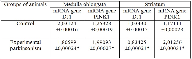
Note: * - reliability relative to control p <0,05
Namely, a decrease in the level of mRNA expression of the DJ1 gene: in the medulla oblongata - by 11.4%; in the striatum - by 19.2%, but the dynamics of change of mRNA expression of the PINK1 gene level was reversed to that established relative to the DJ1 gene. The increase in PINK1 gene expression was in the medulla oblongata - 59.2%; in the striatum - 71.8% (Fig. 1).
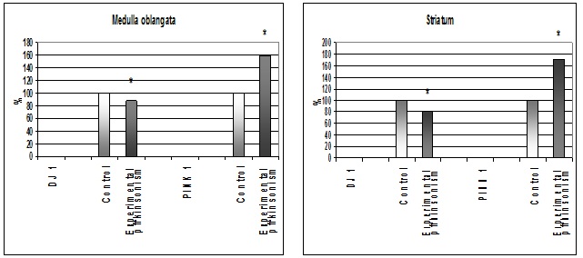
Figure 1 - Changes in mRNA expression of DJ1 and PINK1 genes under experimental parkinsonism. * - reliability relative to control p <0,05.
Because DJ1 is thought to be directly involved in the development of mitochondrial dysfunction, is a sensor of oxidative stress and is able to eliminate peroxide compounds by self-oxidation, and PINK1 acts as a sensor of mitochondrial damage and promotes this process with significant accumulation, it can be stated that the previously found structural and functional damage to organelles in PD are genetically determined and occur largely due to depolarization of mitochondrial membranes, in violation of protein imports and increased sensitivity to peroxide compounds.
Therefore, the changes we found in the mitochondrial apparatus of brain tissue cells may have a genetic component. It was found that under the influence of rotenone there is a significant increase in the number of structurally damaged mitochondria in the medulla oblongata in the absence of significant changes in their total number. The number of structurally damaged organelles was more than 40% (Fig. 2).
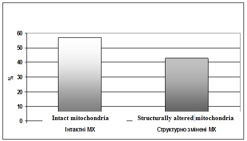
Figure 2 - The ratio of the ultrastructure of mitochondria under experimental parkinsonism.
At the same time, in the striatum with the administration of rotenone there is a restoration of dynamic processes in the mitochondrial apparatus, namely the activation of the process of fission-fusion of mitochondria, which is probably have the compensatory-adaptive character (Fig. 3).

Figure 3 - Ultrastructure of the mitochondrial apparatus of the striatum with the administration of rotnon. MX - mitochondria, f-fMX - fission-fusion process of mitochondria. x 9600
Thus, it can be assumed that in the brain tissues is realized both the ability of DJ1 to be a sensor of oxidative stress and eliminate peroxides by self-oxidation, and the ability of PINK1 to act as a sensor of mitochondrial damage and even promote this process. Moreover, these processes are tissue-specific. To elucidate the exact mechanisms of such processes, further study of structural-functional and genetically determined changes in tissues under experimental parkinsonism is necessary.
Conclusions.
It has been shown that under experimental parkinsonism there is a decrease in the level of mRNA expression of the DJ1 gene in the medulla oblongata and striatum.
It was found that under experimental parkinsonism there is an increase in PINK1 gene expression in both the medulla oblongata and the striatum.
It has been shown that under experimental parkinsonism there are significant changes in the mitochondrial apparatus in the cells of the medulla oblongata and striatum, which have a tissue-specific character and are probably associated with changes in the intensity of mRNA expression of the studied genes.
References.
1. Petrishchev NN, Vlasov TD. Physiology and pathophysiology of the endothelium. Endothelial dysfunction. - SPb: SPbGMU, 2003. – P. 4-38.
2. Lesage S, Brice A. Parkinson's disease: from monogenic forms to genetic susceptibility factors // Hum Mol Genet. - 2009. – V. 18, N 1. – P. 48-59.
3. Rub C, Wilkening A, Voos W. Mitochondrial quality control by the Pink1. Parkin system // Cell Tissue Res. – 2017. – V. 367, N 1. – P. 111-123.
4. Truban D, Hou X, Caulfield TR, et al. PINK1, Parkin, and mitochondrial quality control: what can we learn about Parkinson's disease pathobiology ? // J Parkinsons Dis. - 2017. – V. 7, N 1. – P. 13-29.
5. Canet-Aviles RM, Wilson MA, Miller DW, et al. The Parkinson's disease protein DJ-1 is neuroprotective due to cysteine-sulfinic aciddriven mitochondrial localization // Proc Natl Acad Sci U S A. – 2004. V. 101, N 24. – P. 9103-9108.
6. Gao J, Wang L, Liu J, et al. Abnormalities of Mitochondrial Dynamics Diseases in Neuroses. - Antioxidants (Basel). 2017 - Internet resource: Access mode - https://www.ncbi.nlm.nih.gov/pmc/articles/PMC5488005/
7. Burbulla1 LF, Song P, Mazzulli1 JR, et al. Dopamine oxidation mediates mitochondrial and lysosomal dysfunction in Parkinson's disease // Science. – 2017. - V. 357, N 6357. - P. 1255-1261.
8. Patten DA, Wong J, Khacho M, Soubannier V, Mailloux RJ, Pilon-Larose K, MacLaurin JG, Park DS, McBride HM, Trinkle-Mulcahy L , Harper ME, Germain M, Slack RS. - OPA1-dependent cristae modulation is essential for cellular adaptation to metabolic demand. - EMBO J. 2014 Nov 18; 33 (22): 2676-91
9. Malinovskaya NA, Gasymly ED, Baglaeva OV, et al. Experimental rotenone models of Parkinsons disease in rats. – Modern problems and ways of their solutions in science, transport, production and education. - 2012. - Internet resource: Access mode - https://www.sworld.com.ua/konfer29/1114.pdf
10. Karupu VYa. Electron microscopy. - К .: Vyshcha shkola, 1984. – 208 p.
11. Weekly B. Electron microscopy for beginners. - M .: Mir, 1975. - 326 p.
12. Weibel ER. Morphometry of human lungs. - M.: Medicine, 1970. - 170 p.
13. Tashke K. Introduction to quantitative cyto-histological morphology. - Bucharest: Izd-vo Akademii SRR, 1980. - 192 p.
14. Dave KD, De Silva S, Sheth NP, et al. Phenotypic characterization of recessive gene knockout rat models of Parkinson's disease // Neurobiology of Disease. – 2014. - V. 70, N 1. – P. 190–203.
15. Michael RR, Melkonyan BH, Thanos S. Life-time expression of the proteins peroxiredoxin, beta-synuclein, PARK7 / DJ-1, and stathmin in the primary visual and primary somatosensory cortices in rats // Frontiers in Neuroanatomy. – 2015. - V. 9, N 1. - P. 190-203.
16. Rezai1 M, Mahmoodi M, Ayat Kaeidi A, et al. Effect of crocin carotenoid on BDNF and ventral gene expression area of morphine treated rats // Asian Pacific Journal of Tropical Biomedicine. – 2018. – V. 8, N 8. – P. 387-393.
17. Osipov VP, Lukyanova EM, Antipkin YG, et al. Methods of statistical processing of medical information in scientific research. - K.: Planet of people, 2002. - 200 p.

Admin- Admin
- Сообщения : 171
Дата регистрации : 2016-05-29 -

Надія Теліус и Oleg23451995 поставили лайк
Re: Sidoryak N.G., Putiy Y.V., Rozova E.V. INVESTIGATION OF STRUCTURAL-FUNCTIONAL CHANGES AND FEATURES OF GENES - CANDIDATE EXPRESSION IN TISSUES OF MEDULLA OBLONGATA AND STRIATUM UNDER EXPERIMENTAL PARKINSONISM
автор игор пчела Ср Янв 20, 2021 7:08 am
Good morning!
Thank you for such an interesting article! What are the implications of your findings for the treatment of humans? In your opinion, is there a chance that healthcare providers will be able to deter the progression of cognitive deterioration one day?
Best Regards
Thank you for such an interesting article! What are the implications of your findings for the treatment of humans? In your opinion, is there a chance that healthcare providers will be able to deter the progression of cognitive deterioration one day?
Best Regards
игор пчела- Сообщения : 3
Дата регистрации : 2021-01-19
Re: Sidoryak N.G., Putiy Y.V., Rozova E.V. INVESTIGATION OF STRUCTURAL-FUNCTIONAL CHANGES AND FEATURES OF GENES - CANDIDATE EXPRESSION IN TISSUES OF MEDULLA OBLONGATA AND STRIATUM UNDER EXPERIMENTAL PARKINSONISM
автор Юлия Чт Янв 21, 2021 4:11 pm
Sorry by my question, but.... I can't anderstand.... Where you take the bio-material for your experience?
Юлия- Сообщения : 36
Дата регистрации : 2017-05-26
игор пчела поставил(а) лайк
Интернет-конференция - 2020 :: Секція 2. Фізична реабілітация, Фізична терапія, Ерготерапія, Фізичне виховання. Інклюзивна освіта
Страница 1 из 1
Права доступа к этому форуму:
Вы не можете отвечать на сообщения|
|
|